


Congenital hydrocephalus is typically recognized in patients 2 to 3 month of age, the earlier it is diagnosed the better.
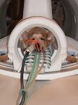
Diagnosis of hydrocephalus is suspected based on the physical examination and signalment (description by peculiar, appropriate, or characteristic marks). It may be suspected based on age, breed, physical appearance and symptoms. However, a definitive diagnosis requires advanced imaging like MRI, CT Scan or ultrasound. If there is an open fontanelle, then ultrasound may be used to see through the skull and measure the size of the lateral ventricles, which are the large chambers in the brain that hold the CSF. Ultrasound does not require anesthesia or sedation for most patients. For CT or MRI, the patient must be under general anesthesia. A patient with hydrocephalus has enlarged ventricles and a smaller amount of cerebral cortex compared to a normal puppy. A CT or MRI will also show if there is any other reason for obstruction to flow of the CSF, such as a tumor. Radiographic evidence (x-ray) suggestive of hydrocephalus includes doming of the calvarium with thinning of cortical bone, decreased prominence of normal calvarial convolutions, and persistent fontanelles. Diagnosis is better assisted by ultrasonography through the fontanelles and confirmed with advanced imaging.
MRI Day


|
|


|
|
No comments:
Post a Comment